|
|
|
|
|
| Available: 2 |
| List Price:$400.00 |
|
|
| Edition: | 1st |
| Publisher: | Thieme Medical Publishers, Incorporated |
| ISBN: | 1-62623-986-X (162623986X) |
| ISBN-13: | 978-1-62623-986-9 (9781626239869) |
| Binding: | Hardcover |
| Copyright: | 2026 |
| Publish Date: | 10/26 |
| Volume: | 4 |
| Weight: | 9.22 Lbs. |
| Pages: | 1322 |
| Subject Class: | RAD (Radiology) |
| Return Policy: | Returns accepted up to 12 months provided no other recalls or return restrictions apply. |
|
|
| |
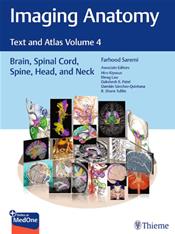
| Abstract: | A visually rich radiologic guide to the brain, spinal cord, head, and neck structuresNormal imaging anatomy and variants, including diagnostic and surgical anatomy, are the cornerstones of radiologic knowledge. Imaging Anatomy: Text and Atlas Volume 4; Brain, Spinal Cord, Spine, Head, and Neck is the fourth in a series of richly illustrated radiologic references edited by distinguished radiologist Farhood Saremi. The atlas is coedited by esteemed colleagues Hiro Kiyosue, Meng Law, Dakshesh Patel, Damin Snchez-Quintana, and R. Shane Tubbs with contributions from an impressive cadre of international authors. Succinctly written text and superb images provide readers with a virtual, user-friendly dissection experience.This exquisitely crafted atlas combines fundamental core anatomy principles with modern imaging and post-processing methods to increase understanding of intricate anatomical features. The book features 36 chapters, several of which are available exclusively online. The chapters cover terminology, classification, and detailed anatomic imaging of the brain, spinal cord, head, and neck structures, along with discussion of relevant associated components for each anatomical area.Key HighlightsHigh-quality cross-sectional multiplanar and volumetric color-coded CT, MRI, and angiography imaging techniques provide detailed insights on specific anatomyCross-sectional and topographic cadaveric views by internationally known anatomists, including the late Albert L. Rhoton Jr., coupled with more than 2,500 illustrations elucidate complex anatomical conceptsConsistently formatted chapters include an introduction, embryology, review of anatomy, discussion of anatomical variants, surgical anatomy, and congenital and acquired pathologiesThis unique resource provides an excellent desk reference for differentiating normal versus pathologic anatomy. It is essential reading for medical students, radiology and neurosurgery residents, neuroradiologists, and neurosurgeons.This print book includes a scratch off code to access a complimentary digital copy on MedOne.Publishers Note: Products purchased from Third Party sellers are not guaranteed by the publisher for quality, authenticity, or access to any online entitlements included with the product. |
|
|
|
|
|
|
Follow Matthews Book Co. on:

Copyright © 2001-2026 Matthews Book Company - All rights reserved. - 11559 Rock Island Ct., Maryland Heights, MO, 63043 - (800) MED-BOOK
Matthews